
D I G I T A LD E N T I S T R Y
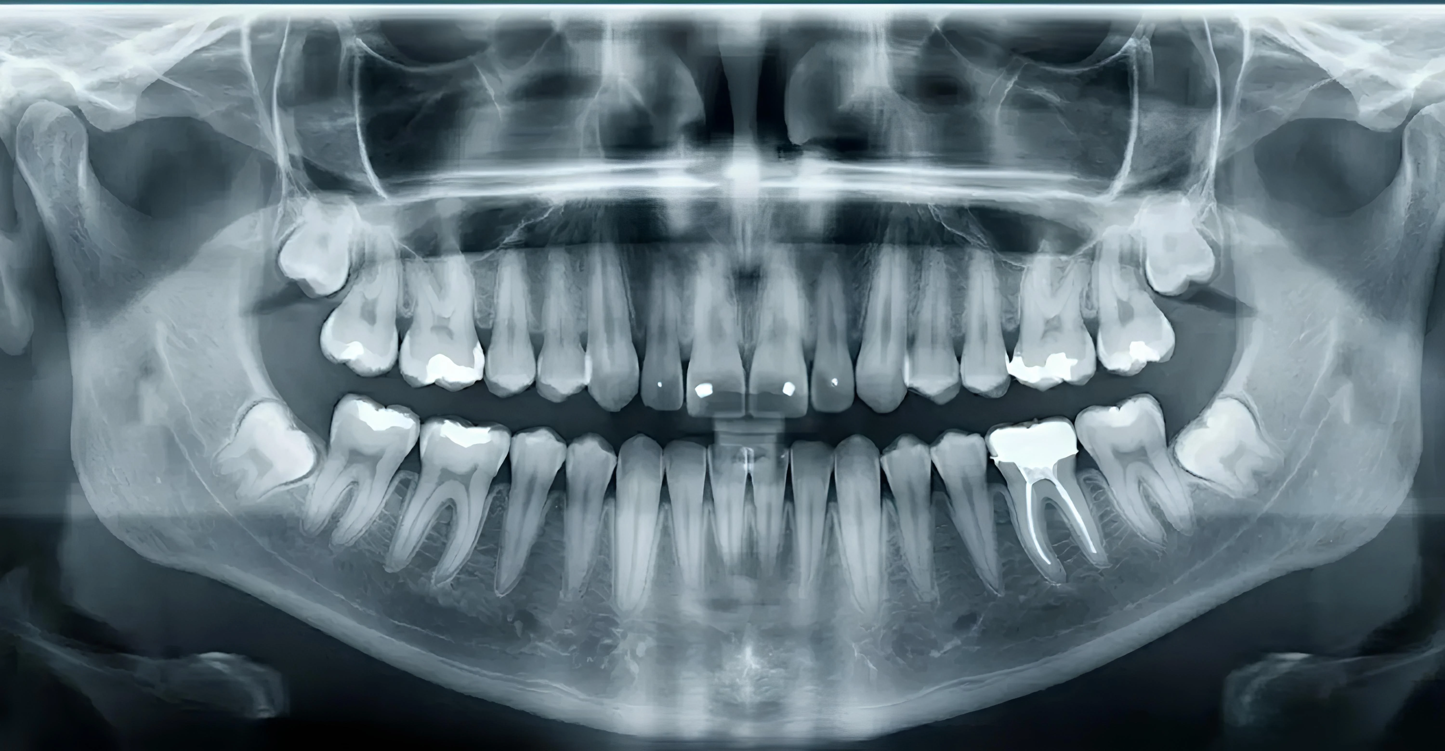
Digital 3D Diagnostics
State-of-the-art diagnostic technology in Sheptytskyi. Low-dose Cone Beam Computed Tomography (KaVo 3D eXam) and digital intraoral optical impressions (Dentsply Sirona Primescan) for comfortable, stress-free care.
KaVo 3D eXam: High-Definition 3D CBCT
The gold standard in maxillofacial 3D X-ray diagnostics, delivering ultra-low radiation doses and rapid 8.5-second scan cycles.

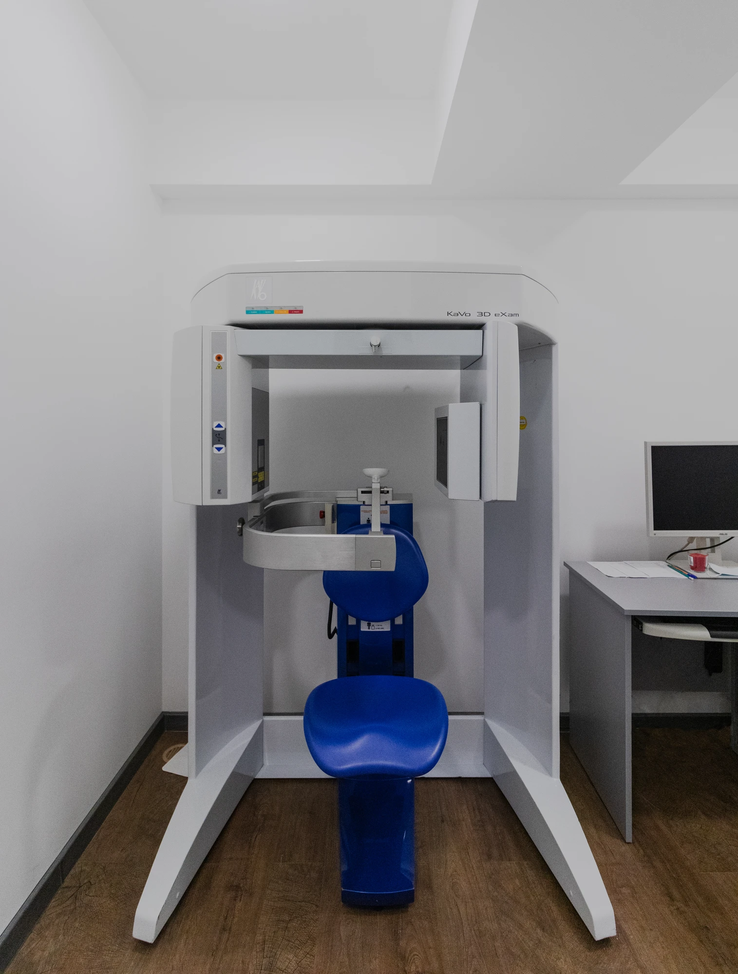
Safe 3D diagnostics from your first visit
The KaVo 3D eXam cone beam computed tomography system is the global gold standard in maxillofacial diagnostics. Pulsed radiation minimizes exposure, ensuring safe examination for adults and children.
Ultra-low radiation dose
Pulsed cone-beam technology reduces radiation exposure significantly compared to conventional medical spiral CTs, ensuring safe imaging for both adults and children.
Comfortable seated examination
The patient is comfortably seated in a cushioned chair within an open-room setting. Zero claustrophobia, zero enclosed tunnels — quick and entirely stress-free.
Rapid 8.5-second scan
A comprehensive 3D volume of both jaws, joints, and maxillary sinuses is captured in just seconds with a single smooth rotation.
High-definition voxel (down to 0.125 mm)
Reveals hidden root canals, micro-fractures, periodontal bone levels, and precise mandibular nerve paths with micrometer clarity.
Interactive 3D CT Reconstruction (Volume Rendering)
CBCT 3D ReconstructionThree-dimensional surface reconstruction of bone and dental structures captured on the KaVo 3D eXam. Rotate, zoom, and inspect the anatomy in full 3D space.
Dentsply Sirona Primescan: Digital Optical Impressions
The fastest and most precise intraoral scanner in modern dentistry. Completely eliminates uncomfortable impression trays and pastes.
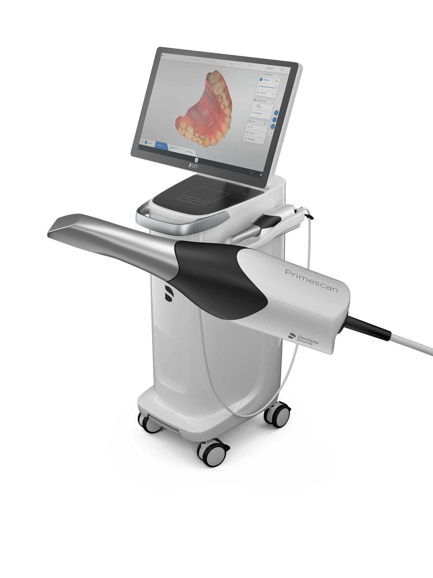
Comfortable optical impressions in 60 seconds
The Dentsply Sirona Primescan captures sub-micron optical impressions without bulky impression trays or gag reflexes. Smart Pixel technology processes over 1,000,000 3D points per second, streaming digital meshes directly to the CAD/CAM lab.
Zero gag reflex, no impression paste
No cold, bulky silicone or alginate trays. The slim optical wand scans both dental arches smoothly in under 60 seconds.
Over 1,000,000 3D points per second
The Smart Pixel sensor captures sub-micron surface details, margin lines, and subgingival tooth anatomy with unmatched fidelity.
Instant CAD/CAM lab transfer
Digital STL models are sent directly to dental technicians in seconds, significantly accelerating crown, veneer, and aligner turnaround.
Flawless marginal restoration fit
Eliminates material shrinkage and distortion errors, ensuring your crown or veneer fits perfectly on the very first try.
Interactive 3D Lower Jaw Intraoral Scan (STL)
Optical STL ScanHigh-resolution optical digital scan of teeth and gingival tissues. Rotate and examine occlusal morphology and preparation margins.
Download Original (.STL, 17.8 MB)Dürr Dental VistaRay 7: High-Precision CMOS Radiovisiography
Next-generation German intraoral digital X-ray system with an ultra-sensitive CMOS sensor. Instant high-definition diagnostics directly at the dental chair with minimal radiation exposure.
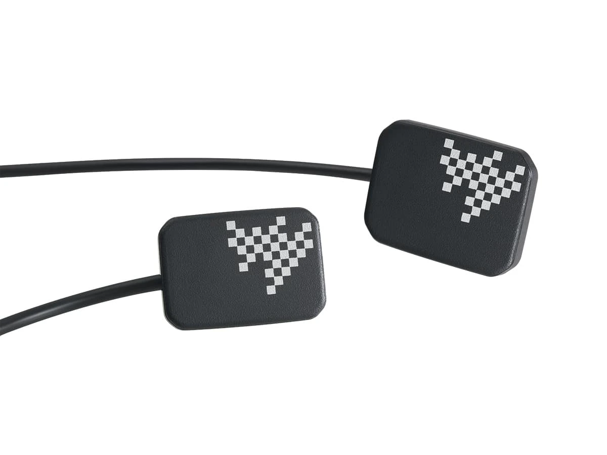
High-Sensitivity CMOS
Next-generation silicon detector delivers maximum contrast for enamel, dentin, and delicate root canals.
Over 25 lp/mm Resolution
Theoretical resolution up to 40 lp/mm reveals the finest hairline cracks and lateral canal branches.
Image in Under 2 Seconds
Instant digital image display directly on the chairside doctor monitor without interrupting treatment.
Up to 90% Lower Radiation
Superior quantum efficiency minimizes patient radiation exposure, ensuring complete clinical safety.
Sample Intraoral Radiographs from CMOS Sensor
DÜRR DENTAL • 3 SCANS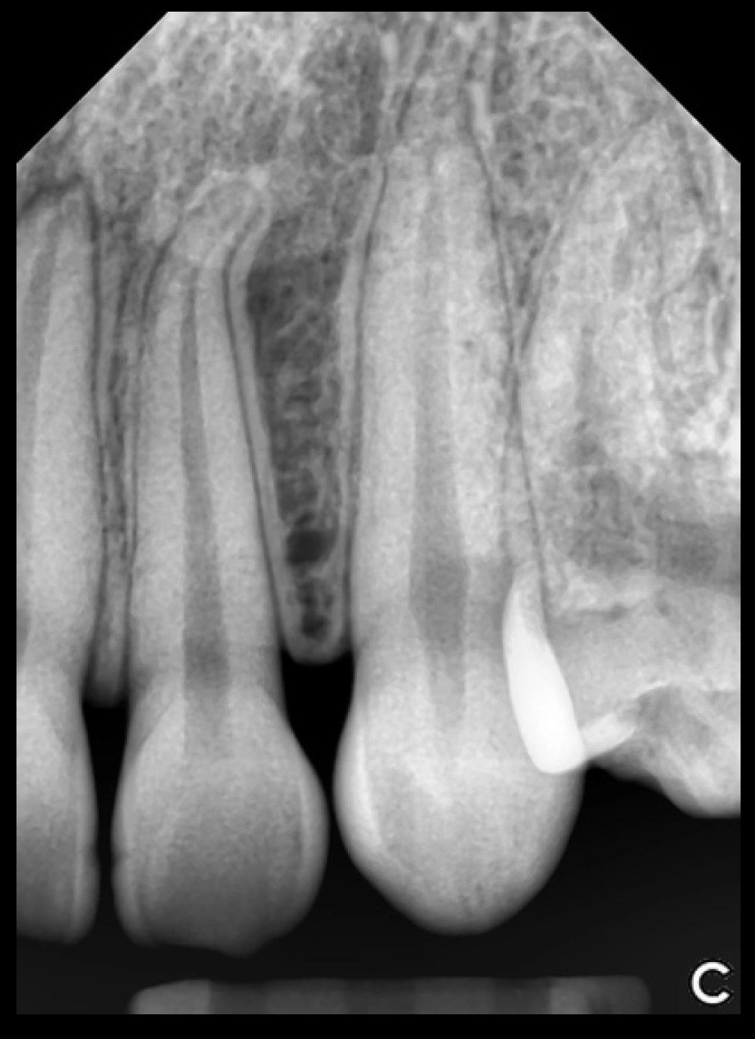
Root Canal & Apex Verification
Assessment of exact working length, root canal morphology, and dense obturation sealing up to the apex.
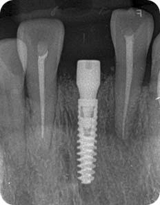
Implant Placement Control
Targeted evaluation of implant positioning, abutment seating, cortical anchorage, and peri-implant bone stability.
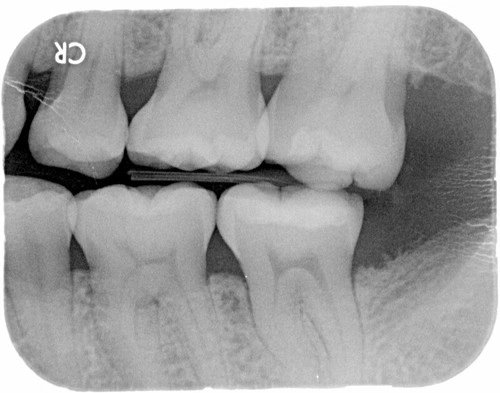
Periodontal Bone, Ligament & Caries
Comprehensive assessment of periodontal ligament space, cortical bone density, and hidden interproximal caries detection.
The Vital Role of 3D Diagnostics Before Treatment & Implantation
A fully digital clinical workflow ensures 100% predictable outcomes, prevents complications, and shortens surgical time.
Virtual implant positioning
The surgeon pre-plans the exact angulation, diameter, and depth of every implant in specialized 3D software prior to surgery.
Complete nerve & sinus protection
Precise 3D measurements eliminate risks to the inferior alveolar nerve and allow accurate planning of sinus lifts when bone height is reduced.
Custom 3D-printed surgical guides
Individual navigation guides printed from 3D models direct surgical drills with sub-millimeter precision.
Long-term implant longevity
Optimized occlusal load distribution prevents bone resorption and secures predictable osseointegration for decades.
Interactive 3D Surgical Implantation Model (GLB)
Surgical Plan GLBComprehensive 3D model of implant placement within mandibular bone architecture. Explore bone depth, fixture angles, and structural support.
Book a consultation
The clinic takes appointments by telephone only. Call us and we will find you a time.



Appointments by phone only